
Fig. 7.
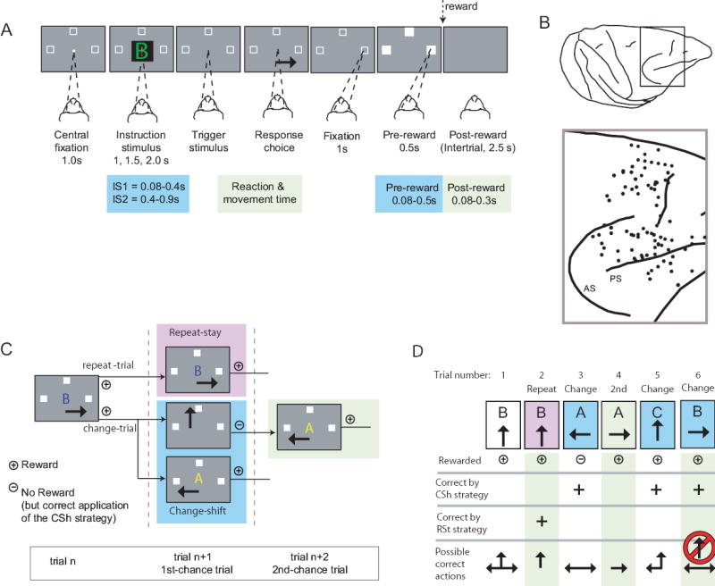
Cell preferring the mapping task. This neuron was located in PFdm (see Fig. 1B). A, B and C each show neuronal activity relative to the onset of the instruction stimulus. Neuronal activity averages: red for the mapping task, black for the strategy task. Change trials differ (p<0.05, Mann-Whitney U Test) but repeat trials do not (p=0.49). D. Three ISs, with arrows indicating the correct action for each. E. Percent of cells by task period showing a task effect, for each monkey. Abbreviation: rew, reward.