Figure 1.
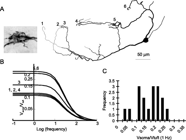
AOB mitral cell tufts are electrically remote from the soma. A, Reconstruction of an AOB mitral cell having six tufts terminating in five glomeruli. The inset shows convergence between tufts 2 and 3. B, Data from simulations showing the decay of voltage changes from tuft to soma as a function of the frequency of a sinusoidal input (current injection). Each line represents the voltage attenuation from a single tuft of the reconstructed cell shown in A plotted against the frequency of the sine wave. C, Histogram of passive attenuation from tuft to soma for 17 tufts from four reconstructed cells simulated in Neuron.

