
Fig. 1.
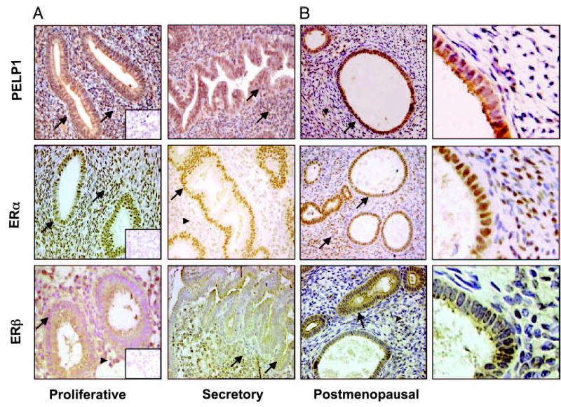
Immunohistochemical localization of ERα , ERβ, and PELP1 in various stages of benign endometrium. A, PELP1 immunostaining was high in glandular and stromal cells (arrow) during the proliferative and secretory phases of benign endometrium. High ERα staining was observed in the glandular cells of the proliferative and secretory phases. In stromal cells, ERα staining was high in the proliferative phase (arrow) and decreased in the secretory phase (arrowhead). Prominent staining of ERβ in the glandular cells was observed in both the proliferative and secretory phases (arrow). In stromal cells, ERβ expression was less prominent in the proliferative phase (arrowhead) and increased in the secretory phase (arrow). Insets (magnification, ×200) show negative controls. B, In postmenopausal endometrium, PELP1 staining was observed only in the glandular compartment (arrow), strong ERα staining in both the glandular and stromal cells (arrow), and strong ERα staining in the glandular cells (arrow) and weak staining in stromal cells (arrowhead) (magnification, ×200). Higher magnification of PELP1, ERα, and ERβ expression in postmenopausal sections is also shown (far right).