
Figure 1.
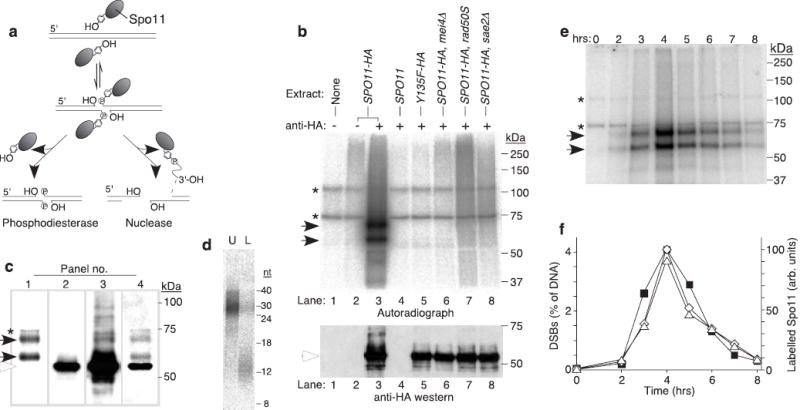
Endonucleolytic processing of covalent Spo11-DSB complexes. a, Alternative mechanisms for Spo11 release1. b, Detection of Spo11-oligonucleotide complexes. Immunoprecipitates from the indicated genotypes with or without antibody were labelled with TdT. Upper panel, autoradiograph; lower panel, anti-HA western. c, Relative mobilities of free and oligonucleotide-associated Spo11-HA. Panel 1, autoradiograph; panels 2–3, low and high exposures of an anti-HA western; panel 4, re-exposure of the blot to film after partial fading of the chemiluminescent signal. d, Sizes of Spo11-associated oligonucleotides from the upper (U) and lower (L) SDS-PAGE bands. Size standards are indicated. e, Time course of appearance of Spo11-oligonucleotide complexes during meiosis. f, Quantification of upper (⋄) and lower (Δ) labelled Spo11 species from e, and the DSB frequency at the HIS4LEU2 recombination hotspot measured in the same culture (▪). Each point is a single measurement. Asterisks, non-specific labelling; closed arrows, Spo11-specific labelled species; open arrows, free Spo11-HA.