
Figure 1.
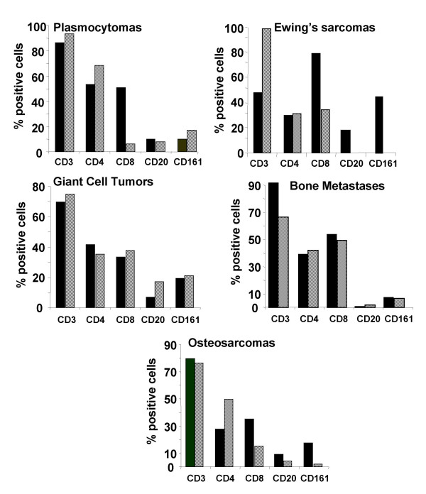
Phenotypic analysis of TIL isolated from human bone associated tumors compared with the phenotype of autologous PBL. TIL were isolated from tumor biopsies and cultured for 21 days in RPMI 1640 supplemented with 10% FCS and 300 UI/ml IL-2. Autologous PBL were isolated from peripheral blood samples after gradient density centrifugation and cultured in the same conditions for 21 days. After the indicated culture period, the phenotype of TIL (hatched bars) and PBL (dark bars) were analyzed by flow cytometry. The percentages of positive cells are given as median value. Plasmocytoma, n = 2; Ewing's sarcoma, n = 2; giant cell tumor, n = 7; bone metastase, n = 4; osteosarcoma, n= 6.