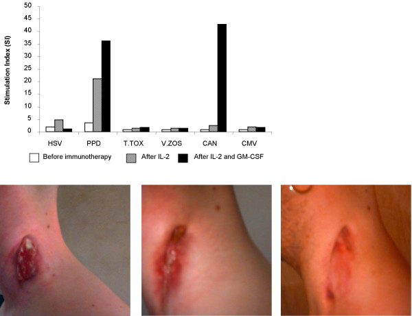
Figure 2.
(a – top) Specific lymphoproliferative responses to recall antigens of patient 4. Data are at IRIS presentation (white bars), 4 weeks after IL-2 administration (hatched bars) and 4 weeks post final IL-2 and GM-CSF dosing (solid bars). Photographs depict the clinical manifestation of MAC lymphadenitis of the neck in patient 4, at IRIS presentation (b – bottom left), 4 weeks after IL-2 administration (c- bottom centre), and 4 weeks after IL-2 plus GM-CSF administration (d – bottom right).

