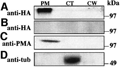
Fig. 3. HET-C is in the plasma membrane fraction. (A) CJ44 (het-cPA::HA) compatible transformants were subjected to cell fractionation and anti-HA-aminolinked column chromatography. Eluates from cellular fractions were probed by western analysis with a monoclonal anti-HA antibody. (B) CJ44 (het-cPA) compatible transformants treated identically to (A) show lack of cross-reacting material. (C) Western analysis using antibodies to the PM H+-ATPase (Bowman et al., 1981) show the predicted ∼100 kDa PM H+-ATPase band. (D) Western analysis using antibodies to β-tubulin, a cytoplasmic marker. MW markers are indicated. PM, plasma membrane fraction; CT, cytoplasmic fraction; CW, cell wall fraction.
