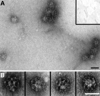
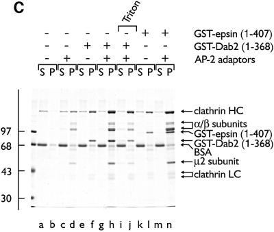
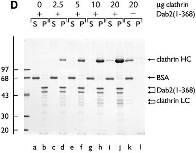
Fig. 8. Dab2 co-operates with AP-2 to drive the assembly of invaginated clathrin buds upon lipid membranes. Clathrin was added to 10% PtdIns(4,5)P2-containing lipid monolayers pre-incubated in the absence (A, inset) or presence (A and B) of GST–Dab2(1–368). An EM grid was used to remove each monolayer and then negatively stained. Bar = 50 nm. (C) Phosphoinositide-containing liposomes were first pre-incubated with AP-2 (lanes c, d, g–j and m, n), GST–Dab2(1–368) (lanes e–j) or GST–epsin 1(1–407) (lanes k–n) at 4°C for 60 min as indicated. After recovery by centrifugation, each liposome pellet was resuspended and then incubated at 4°C for 60 min with purified clathrin trimers in the presence of carrier BSA. After centrifugation, aliquots of 1/40 of each supernatant (S) and 1/4 of each pellet (P) were resolved by SDS–PAGE and stained with Coomassie Blue. Before centrifugation, Triton X-100 (1% final) was added in one reaction (lanes i and j). (D) PtdIns(4,5)P2-containing liposomes were first pre-incubated with (lanes a–j) or without (lanes k and l) thrombin-cleaved Dab2(1–368) at 4°C for 60 min. After recovery by centrifugation, each liposome pellet was resuspended and then incubated at 4°C for 60 min with increasing amounts of purified clathrin trimers in the presence of carrier BSA. After centrifugation, aliquots of 1/25 of each supernatant (S) and 1/5 of each pellet (P) were resolved by SDS–PAGE and stained with Coomassie Blue.

An official website of the United States government
Here's how you know
Official websites use .gov
A
.gov website belongs to an official
government organization in the United States.
Secure .gov websites use HTTPS
A lock (
) or https:// means you've safely
connected to the .gov website. Share sensitive
information only on official, secure websites.


