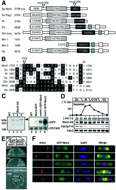
Fig. 1. Identification of Mor2. (A) A schematic presentation of the homology between Mor2 and Furry homologous proteins. Schizosaccharomyces pombe Mor2 is aligned with S.cerevisiae Pag1/Tao3 (Sc), Arabidopsis thaliana T51397 (At), C.elegans AAF99910 (Ce), D.melanogaster Furry (Dm), Mus musculus CAC42175.1 (Mm1), CAC42196.1 (Mm2) and Homo sapiens CAB42442 (Hs). Identities (%) and the number of amino acids are indicated. (B) The mutation site of the mor2-282 allele. (C) Identification of Mor2 protein. Lanes 1–4: total cell extract prepared from wild type (WT, 972), and the cells having mor2+:3HA:kanr (Mor2-HA, YS78-1) grown in YPD medium were electrophoresed on SDS–PAGE gels and immunoblotted with anti-HA (lanes 1 and 2). Total cell extracts (T, lane 2) prepared from the Mor2-HA strain were fractionated into soluble (S, lane 3) and insoluble fractions (P, lane 4) by centrifugation at 14 000 g. Lanes 5–8: wild type, and cells with mor2+:GFP:kanr (Mor2–GFP, YS77), kanr:nmt41-GFP:mor2+ (nmt41–GFP–Mor2, YS84-1) or kanr:nmt1-GFP:mor2+ (nmt1-GFP–Mor2, YS85) grown in Edinburgh minimal medium (EMM) containing 4 µM thiamine were transferred into EMM. After cultivation for 14 h, the cells were collected. Total cell extracts prepared from the cells were immunoblotted with anti-GFP or anti-PSTAIR antibodies. (D) Mor2 protein during the cell cycle. Early G2 cells of Mor2-HA strain were collected by centrifugal elutriation and cultured in YPD medium at 28°C. Samples of the culture were taken at the indicated times for calculating the percentage of septated cells. Total cell extracts prepared from the samples of the same cultures were immunoblotted with anti-HA (for Mor2-HA), anti-PSTAIR (for Cdc2) or anti-Cdc2 phosphorylated on tyrosine-15 (for Cdc2-P) antibodies. (E) Growth (upper) and morphology (lower) of the Mor2-over-expressing cells. Upper panel: the nmt1-GFP–Mor2 and nmt41–GFP–Mor2 strains were cultured on EMM (ON) or EMM plates containing 4 mM thiamine (OFF) at 25°C for 3 days. Lower panel: the nmt1-GFP–Mor2 strain grown in EMM containing 4 µM thiamine was transferred into EMM medium and cultured for 18 h at 25°C. (F) Localization of Mor2 and F-actin. The nmt41–GFP–Mor2 strain cells grown in EMM containing 4 µM thiamine were transferred into EMM medium. After cultivation for 14 h, cells were fixed and stained with rhodamine– phalloidin. Merged image (Merge): F-actin (red), GFP–Mor2 (green) and DAPI (blue).

An official website of the United States government
Here's how you know
Official websites use .gov
A
.gov website belongs to an official
government organization in the United States.
Secure .gov websites use HTTPS
A lock (
) or https:// means you've safely
connected to the .gov website. Share sensitive
information only on official, secure websites.
