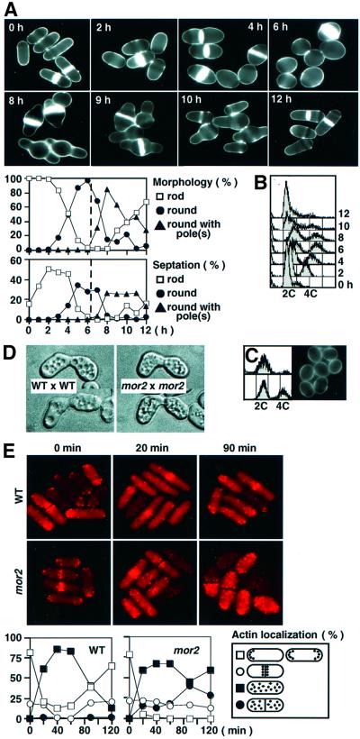
Fig. 3. Phenotypes of the mor2 mutant. (A and B) Reversible phenotype of the mor2 mutant. mor2 mutant cells (DH107-4C) grown in YPD medium at 25°C were transferred to 36°C (time 0 h). After incubation for 6 h, the cells were shifted down to 28°C and incubated for 6 h (total 12 h). The cells at the indicated times were taken for observation of cell morphology after having been stained with Calcofluor (A) and for FACS analysis (B). (C) mor2 mutant cells (DH107-4C) grown in YPD medium at 25°C were transferred to 36°C and incubated for 4 h. The unseptated round cells in the culture were collected by centrifugal elutriation for FACS analysis (upper panels, the collected round cells; lower panels, the culture) and for observation of morphology (isolated round cells). (D) Shmoo morphology of the mor2 mutant. Wild-type (h–) or mor2 mutant cells (h– mor2-786) were crossed with wild-type (h+) or mor2 mutant cells (h+ mor2-786), respectively. After incubation for 24 h on malt extract (MEA) plate, the shmoo morphology of the cells was observed. (E) F-actin localization in the mor2 mutant. The wild-type (972) and mor2 mutant cells (DH192-1A) grown in YPD medium at 25°C were transferred to 36°C (time 0 h). The cells were taken at the indicated times for observation of F-actin localization after being stained with rhodamine–phalloidin.

An official website of the United States government
Here's how you know
Official websites use .gov
A
.gov website belongs to an official
government organization in the United States.
Secure .gov websites use HTTPS
A lock (
) or https:// means you've safely
connected to the .gov website. Share sensitive
information only on official, secure websites.
