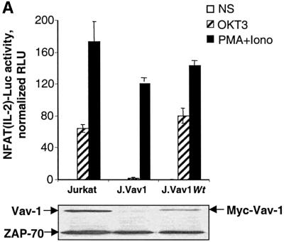
Fig. 3. TCR-dependent NFAT activation. (A) Cells were transfected with 5 µg of pNFAT(IL2)-Luc reporter plasmid, together with the control pRL-TK plasmid. The J.Vav1WT subline was derived by stable transfection of J.Vav1 cells with a wild-type (Wt) Vav-1 expression plasmid. At 18 h post-transfection, the cells were stimulated for 6 h with the indicated agents. The pNFAT(IL2)-Luc activity measured in each sample was normalized to the Renilla luciferase activity to control for transfection efficiency. Bars represent the mean ± standard deviation from triplicate samples. The lower panel indicates the level of Vav-1 protein in each cell population, as determined by immunoblotting. (B) J.Vav1 cells were co-transfected with the pNFAT(IL2)-Luc reporter, plus 10 µg of either wild-type Vav-1 or Vav-1 CH– plasmid DNA. Bars represent the mean luciferase activities from duplicate samples, after normalization to the maximal response obtained with ionomycin plus PMA. The inset shows the expression levels of the FLAG-tagged Vav proteins.

