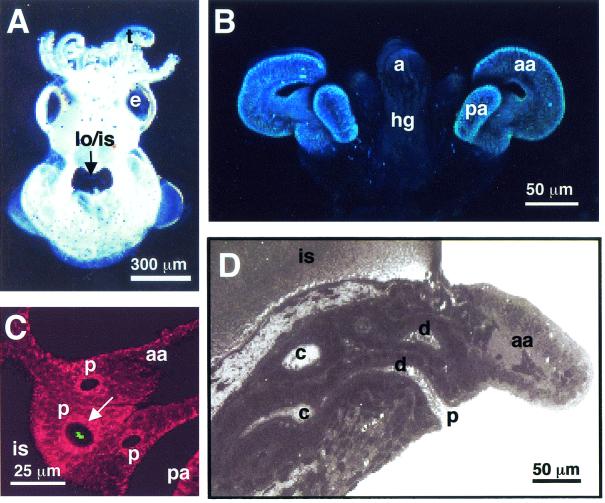
FIG. 1.
Path of symbiont cells into the juvenile host light organ. (A) Hatchling juvenile squid contain a bilobed light organ that is part of the ink sac complex located in the center of the mantle cavity (arrow). (B) Confocal image of the nascent light organ, revealing the ciliated epithelial fields on either side of the light organ, each with two appendages that direct seawater containing environmental bacteria towards sites of colonization. (C) Confocal image showing an aggregation of GFP-labeled V. fischeri (green, arrow) entering one of the three pores that occur on each side of the light organ. The cells of the light organ are stained with CellTracker (red), which is a general stain for eukaryotic cell cytoplasm. (D) Each pore leads to a ciliated duct that terminates in an epithelium-lined crypt space where permanent colonization by the symbiont population takes place. a, anus; aa, anterior appendage; c, crypt space; d, duct; e, eye; hg, hind gut; is, ink sac; lo, light organ; p, pore; pa, posterior appendage; t, tentacle.

