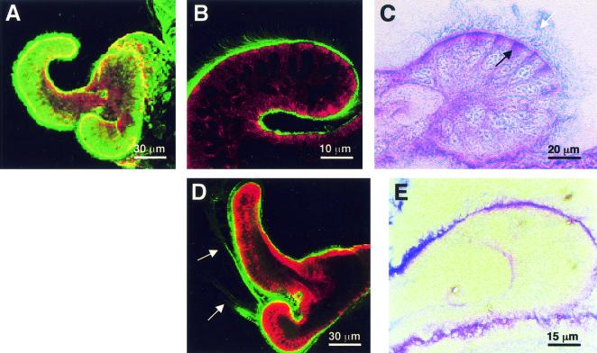
FIG. 2.
Mucin storage and secretion in the superficial ciliated epithelium of the E. scolopes light organ. (A) Confocal image of the light organ surface of a representative live hatchling squid, showing uniform staining with WGA (green) for N-acetylneuraminic acid (sialic acid) residues characteristic of sialomucins. (B) WGA staining of a confocal section through the ciliated fields of a live specimen, confirming the presence of a coat of sialomucin (green) on the surfaces. (C) PAS-AB histological staining of a section of hatchling squid, demonstrating that the cytoplasm of the apical regions of the cells comprising the ciliated epithelial appendages contains dense neutral mucin and sialomucin stores (purple, black arrow), while the extracellular surfaces are coated with sialomucin (blue, white arrow). (D) Representative confocal micrograph showing the typical sialomucin shedding event (arrows) that occurred in the surface epithelium within 1 h of exposure to USW, gram-negative or -positive bacteria, or bacterial PGN. (E) After several hours of exposure to environmental bacteria, the abundance of mucins within the appendages decreased compared with the abundance in hatchling animals (panel C). In the confocal images, the host cells are counterstained with CellTracker (red).

