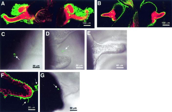
FIG. 3.
Influence of colonization on mucus shedding and bacterial aggregation. (A) Representative confocal micrograph of a 72-h aposymbiotic animal, showing the characteristic thick layer of sialated mucus (green) covering the superficial ciliated epithelial fields. (B) Although a thin coat of sialomucin-containing mucus (green) was present on the outside of the ciliated epithelium, no secretion of mucus was detected by 48 h after successful colonization by V. fischeri. (C) Aposymbiotic animals were capable of aggregating GFP-labeled V. fischeri (arrow) up to 96 h after hatching. (D) Wild-type V. fischeri cells formed loose aggregations (arrow) outside 24-h symbiotic animals. (E) By 48 h, no aggregations of V. fischeri cells were observed outside symbiotic light organs. (F) After the symbionts were removed from the light organ with the antibiotic chloramphenicol, mucus shedding was again detected (green, arrows), although not at the levels seen in aposymbiotic animals that were the same age. (G) The ability to form bacterial aggregations (arrow) was restored in 48-h symbiotic animals that had been cured with antibiotics for 24 h. In the confocal images, the sialomucin is labeled with WGA and the host cells are counterstained with CellTracker (red).

