FIG. 2.
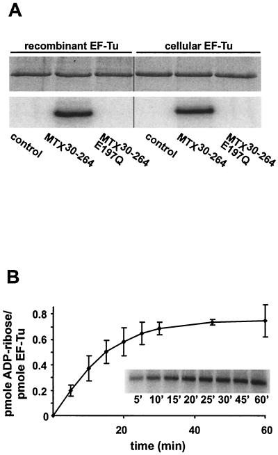
[32P]ADP ribosylation of cellular EF-Tu and recombinant EF-Tu. (A) Cellular EF-Tu and recombinant EF-Tu (2.5 μM) were incubated alone (control) or with MTX30-264 (50 nM) or MTX30-264E197Q (50 nM) in the presence of 100 μM [32P]NAD for 30 min at room temperature. Proteins were separated by SDS-PAGE (upper panel), and labeling was detected by phosphorimaging (lower panel). Quantification of [32P]ADP-ribose incorporation revealed that ∼50% of the total amount of EF-Tu used in this assay was modified. (B) Recombinant EF-Tu (300 nM) was incubated with 100 nM MTX30-264 in the presence of 100 μM [32P]NAD in a total volume of 20 μl for the indicated times. The incorporation of [32P]ADP-ribose was detected after SDS-PAGE by phosphorimaging, and data were quantified with ImageQuant software. The extent of ADP-ribosylation is indicated as picomoles of ADP-ribose incorporated per picomole of EF-Tu. Data are given as means and standard errors (n = 3). The inset illustrates the phosphorimaging data from a representative experiment.

