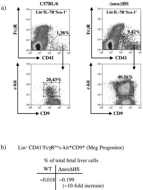
FIG. 1.
Isolation of a Lin− CD41+ FcγRlow c-kit+ CD9+ progenitor cells from mouse E13.5 to E14.5 fetal liver. (a) Flow cytometric analysis of Lin−IL-7R−Sca-1− wild-type and ΔneoΔHS fetal liver cells labeled with CD41-fluorescein isothiocyanate, FcγR-PE, c-kit-APC, CD9-biotin, and streptavidin-APC-Cy7. Sorting gates for isolation of Lin− CD41+ FcγRlow c-kit+ CD9+ cells are shown. (b) The table shows the average frequencies of Lin− CD41+ FcγRlow c-kit+ CD9+ population in wild-type and ΔneoΔHS fetal liver from nine different sorting experiments. Note that only 10% of total fetal liver cells are Lin−.

