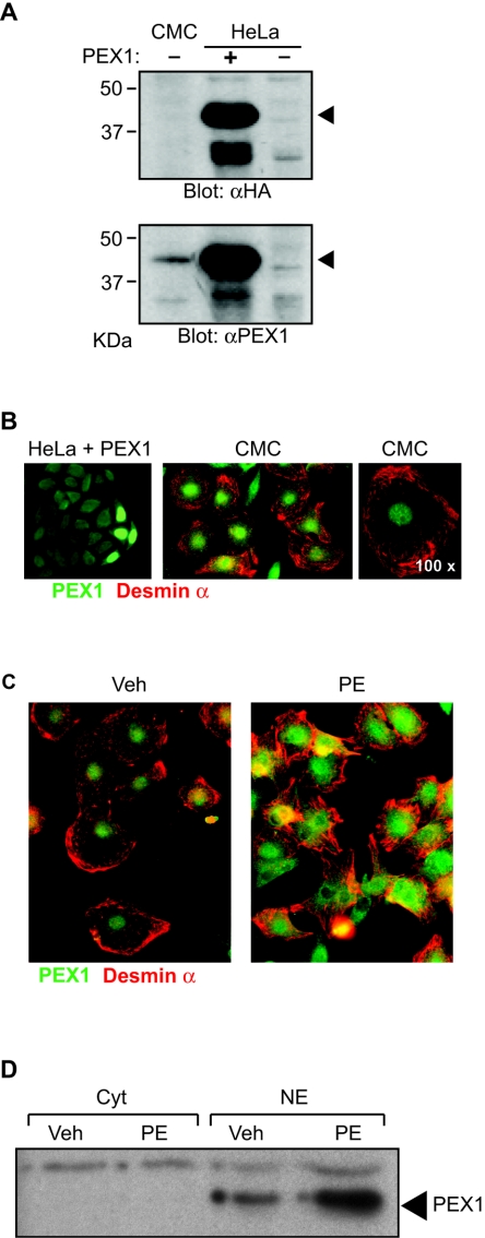
FIG. 5.
Expression and regulation of PEX1 protein in cardiomyocytes. (A) Western blots were generated with nuclear extracts isolated from cardiomyocytes, HeLa cells transfected with HA-tagged PEX1 expression vector, or an empty vector. The membranes were incubated with anti-HA (αHA) or anti-PEX1 (αPEX1) antibodies. (B) PEX1 was detected by indirect immunofluorescence with the anti-PEX1 antibody in HeLa cells transfected with an HA-tagged PEX1 expression vector (left panel) and in postnatal cardiomyocytes. The right panel shows labeling in cardiomyocytes at a 2.5-fold-higher magnification. (C) PEX1 protein level was determined by immunofluorescence in cardiomyocytes treated with vehicle (Veh) or 0.1 mM PE for 48 h. (B) and (C) The cardiomyocytes were costained with an anti-desmin α antibody. (D) The level of PEX1 protein was determined by Western blotting with the anti-PEX1 antibody in nuclear (NE) and cytoplasmic (Cyt) extracts from cardiomyocytes stimulated (PE) or not stimulated (Veh) with 0.1 mM PE for 48 h. CMC, cardiomyocytes.

