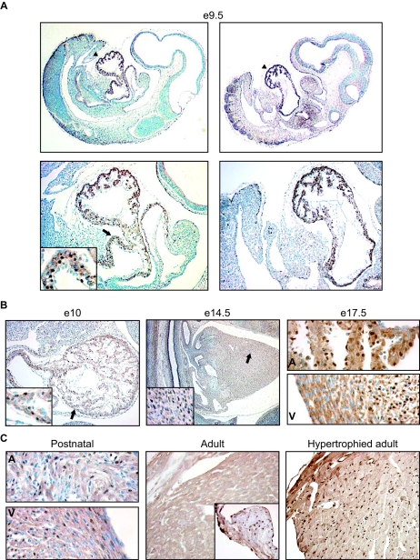
FIG. 6.
Developmental pattern of PEX1 expression in the mouse heart. The expression of PEX1 was determined by immunohistochemistry with the anti-PEX1 antibody on sections from embryos of E9.5, E10, E14, and E17.5 (A and B); from postnatal and adult mouse hearts (C); and from a mouse model of angiotensin II-induced cardiac hypertrophy (38). (A) The two upper panels show expression of PEX1 in the heart in whole embryos. The portion indicated with arrowheads is magnified four times and shown below. (A) and (B) The areas indicated with arrows are magnified four times and shown in the inserts. (C) The insert in the middle panel shows a portion of the aortic valve. A, atria, V, ventricles.

