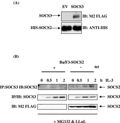
FIG. 7.
SOCS2 and SOCS3 interaction. (A) His-tagged SOCS2 fusion protein (3 μg) was precoupled to 50 μl of 20% nickel-nitrilotriacetic acid beads in protein interaction buffer for 2 h at 4°C. The beads were collected by centrifugation, and lysates from 293T cells transiently transfected with 2 μg empty vector (Ev) or FLAG-tagged SOCS3 were added. Reaction mixtures were incubated for 2 to 4 h at 4°C in protein interaction buffer, washed, and analyzed by SDS-PAGE as described in the text. Membranes were immunoblotted (IB) with anti-FLAG (top) to detect SOCS3 and reprobed with anti-His to detect His-tagged SOCS2 fusion protein (bottom). (B) Ba/F3-SOCS2 cells were grown in the presence and absence of tetracycline for 48 h. Cells were incubated overnight in 2% FCS and stimulated with 100 U/ml IL-3 as shown. Proteasome inhibitors MG132 (0.5 μM) and LLnL (0.5 μM) were added 1 h before lysing. Cell lysates were immunoprecipitated (IP) with SOCS3 antibody and immunoblotted with anti-FLAG (top) or anti-SOCS3 (middle). SOCS2 expression is shown in the bottom panel.

