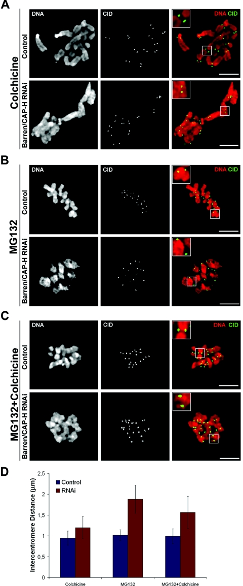
FIG. 6.
Analysis of intercentromere distances after depletion of Barren/CAP-H. (A to C) Both control and Barren/CAP-H-depleted (72 h) cells were immunostained for CID and DNA. Cultures were (A) incubated with 30 μM colchicine for 2 h to depolymerize all microtubules before entering mitosis, (B) incubated for 2 h with 20 μM MG132 to arrest cells in metaphase, (C) incubated with 20 μM MG132 to arrest cells in metaphase followed by a 30-min incubation with 30 μM colchicine to depolymerize all microtubules that were previously attached to the kinetochores. Scale bars are 5 μm. Higher magnifications (2×). (D) Quantification of intercentromere distances of control and Barren/CAP-H-depleted cells after the indicated experimental conditions.

