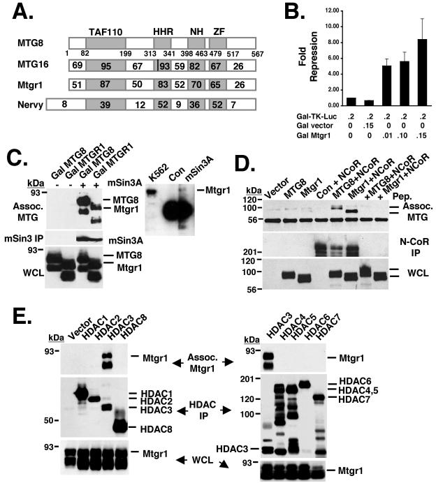
FIG. 1.
Mtgr1 is a transcriptional corepressor. (A) Schematic diagram showing the MTG family members, including Nervy. The number within each box indicates the percent identity of the region compared to MTG8. The vertical line in the HHR domain of MTG16 indicates the two amino acid changes that affect mSin3A binding. TAF110, transcriptional activating factor; HHR, hydrophobic heptad repeat; NH, Nervy homolog; ZF, zinc finger. (B) Mtgr1 repressed the transcription of a heterologous promoter in a dose-dependent fashion. The GAL-TK-luciferase reporter construct was cotransfected into NIH 3T3 cells with a control vector or increasing amounts of pCMV5-GAL-Mtgr1 as shown. Cells were harvested 48 h posttransfection and assayed for luciferase activity. The values shown were normalized with secreted alkaline phosphatase (pCMV-SEAP) as an internal control for transfection efficiency. Numbers shown below the bars are the amounts of DNA in micrograms. (C) FLAG-tagged mSin3A was cotransfected into Cos-7 cells with either empty vector; GAL-MTG8/ETO, used as a positive control (Con); or GAL-Mtgr1. After immunoprecipitation with anti-FLAG, GAL-MTG8 and GAL-Mtgr1 were detected by immunoblotting with anti-GAL4 (top). The expression of mSin3A was confirmed with anti-FLAG after immunoprecipitation (IP) (middle). The expression levels of Gal-MTG8 and Gal-Mtgr1 were confirmed by immunoblotting with anti-GAL (whole-cell lysate [WCL], bottom). In addition, endogenous mSin3A was precipitated with anti-mSin3A and copurifying Mtgr1 was identified by immunoblotting (right). (D) FLAG-N-CoR was cotransfected into Cos-7 cells with either GAL-MTG8 or GAL-Mtgr1. Anti-Gal immunoblots show that both Gal-MTG8 and Gal-Mtgr1 coimmunoprecipitated with N-CoR (top) and are present in the whole-cell lysate (bottom). Addition of the antigenic peptide to the FLAG beads prior to immunoprecipitation was used as a further control (+ Pep.). (E) FLAG-tagged (HDAC1 to -6), HA-tagged (HDAC7), and Myc-tagged (HDAC8) forms of the indicated HDACs were transfected into Cos-7 cells along with GAL-Mtgr1. Immunoprecipitation of the HDACs, followed by Gal immunoblotting (top), was used to detect Mtgr1 associated (Assoc.) with HDAC3. An anti-Gal immunoblot assay of the whole-cell lysate shows that Gal-Mtgr1 was expressed in every lane (bottom), while anti-FLAG/Myc or anti-FLAG/HA immunoblotting indicates that each HDAC was immunoprecipitated (middle) by the FLAG, HA, or Myc antibodies.

