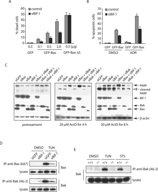
FIG. 7.
Bif-1 contributes to not only Bax, but also Bak activation. (A) siBif-1-expressing HeLa clones or control cells were transfected with the indicated amounts of plasmid DNA encoding GFP, GFP-Bax, or GFP-BaxΔS mutant for 24 h prior to trypan blue dye exclusion assay. The error bars indicate standard deviations. (B) HeLa clones were transfected with 0.5 μg GFP- or GFP-Bax-producing plasmids for 24 h and treated with 1 μM ADR or control DMSO for 8 h. After being stained with DAPI, the apoptotic GFP-positive cells were counted (n > 300). (C) HeLa cells transfected with the indicated siRNAs were treated with 20 μM ActD for the indicated periods of time and subjected to SDS-PAGE/immunoblot analysis with the indicated antibodies. (D) HeLa cells transfected with siGFP or siBif-1 were treated with 4 μg/ml TUN for 24 h prior to the preparation of cell lysates in CHAPS lysis buffer. Immunoprecipitation (IP) was performed with anti-Bak monoclonal antibody (Ab-2) or anti-Bax 6A7 monoclonal antibody, and the resulting immune complexes and total-cell lysates were analyzed by SDS-PAGE/immunoblotting with anti-Bax and anti-Bak polyclonal antibodies. (E) Bif-1−/− and Bif-1+/+ MEFs were treated with 1 μg/ml TUN for 24 h, 1 μM STS for 12 h, or DMSO as a control. Cell lysates were prepared in CHAPS lysis buffer and subjected to immunoprecipitation with anti-Bak monoclonal antibody (Ab-2). The resulting immune complexes, as well as total-cell lysates, were analyzed by SDS-PAGE/immunoblotting with anti-Bak polyclonal antibody.

