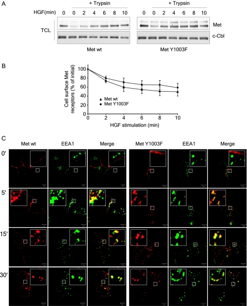
FIG. 5.
Ubiquitination-deficient Met receptor mutant is internalized. (A) T47D cells were stimulated with 3 nM HGF at 37°C and then transferred on ice. Cells were acidified for 10 min and then treated with trypsin for 30 min. Trypsin was inhibited before cells were lysed. Lysates were blotted with Met (DL-21) and c-Cbl antibodies. (B) T47D cells were stimulated with 3 nM HGF at 37°C and then transferred on ice. Cells were acidified for 10 min and then labeled with Met AF276 antibody as described under Materials and Methods. The mean fluorescence intensity per cell was measured by flow cytometry and the percentage of Met receptors remaining at the cell surface over time is plotted. The graph represents the mean ± standard deviation of three independent experiments. (C) Both wild-type Met and Met Y1003F internalize and traffic to early endosomes. T47D cells were plated on coverslips, and 16 h later stimulated with 1.5 nM HGF at 37°C for the indicated times. Coverslips were fixed in 3% PFA and stained with Met AF276 (red) and EEA1 (green) antibodies. Confocal images were taken with a 100× objective and 2× zoom. Yellow staining represents colocalization between Met and EEA1. The bar represents 5 μm.

