FIG. 2.
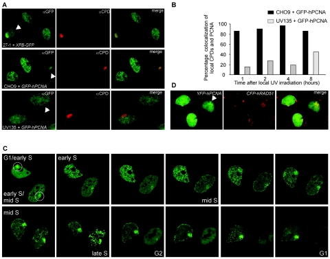
Accumulation of DNA repair proteins at sites of local UV-induced damage. (A) Upper panels: XPB and CPDs colocalize. CHO cells deficient in XPB (27-1) complemented with XPB-GFP were locally UV irradiated (locally damaged cells are indicated with the arrows). One hour after UV irradiation, DNA damage was detected with antibodies against CPDs (shown in red), and XPB-GFP accumulation was detected with antibodies against GFP (shown in green). Colocalization of XPB and CPDs is demonstrated in the merged image. Middle panels: PCNA and CPDs display colocalization. CHO9 cells expressing GFP-hPCNA were locally UV irradiated. One hour after UV irradiation, cells were fixed, DNA was denatured, and DNA damage was detected by indirect immunofluorescence using antibodies against CPDs (shown in red). GFP-hPCNA was also detected by indirect immunofluorescence using antibodies against GFP (shown in green). Shown are three GFP-hPCNA-expressing cells in different stages of the cell cycle. On top of the replication pattern of PCNA, one cell shows local accumulation of GFP-hPCNA, which colocalizes with the CPD signal (shown in yellow in the merged image). Bottom panels: No colocalization of PCNA and CPDs in a NER-deficient cell line, UV135, which is deficient in XPG. Locally irradiated UV135 cells expressing GFP-hPCNA were fixed 1 hour after irradiation and stained using antibodies against GFP (in green) and CPDs (in red). The bottom cell shows local accumulation of CPDs but no accumulation of PCNA. (B) Quantitation of the results shown in Fig. 2A, middle and bottom panels. The percentages of local CPD accumulations that also show local PCNA accumulation after local UV irradiation were determined at the indicated time points for CHO9 and UV135 cells. For all cell lines, 65 to 70 cells were examined. (C) Time-lapse imaging of GFP-hPCNA in cells treated with local UV damage shows transient accumulation of GFP-hPCNA (indicated by the circles) at sites of local UV damage in different stages of the cell cycle. Based on the PCNA replication pattern, the upper cell was at the transition from the G1 phase to the early S phase, and the lower cell was in the early S phase. In the upper cell, this local GFP-hPCNA accumulation disappeared for a short period during the early S phase but reappeared at the transition from the early S phase to the mid-S phase. In the lower cell, the local GFP-hPCNA accumulation disappeared in the G2 phase and the subsequent G1 phase in the daughter cells. Note the decrease in fluorescence signal around mitosis as a result of cells rounding up and detaching from the coverslips. (D) Colocalization of PCNA and Rad51 at site of local damage. Chinese hamster ovary cells stably expressing both YFP-hPCNA and CFP-hRAD51 were locally UV irradiated. Cells were fixed 1, 4, 8, and 16 h after UV irradiation, and local accumulation of PCNA and Rad51 was directly detected using the appropriate filter sets (Chroma) to discriminate between cyan fluorescent protein and yellow fluorescent protein. At all these different time points, we found examples of local accumulations of PCNA (shown in green) and Rad51 (shown in red) during S phase, where the focal pattern of PCNA partly colocalized with the focal pattern of Rad51 (shown in the merged image).

