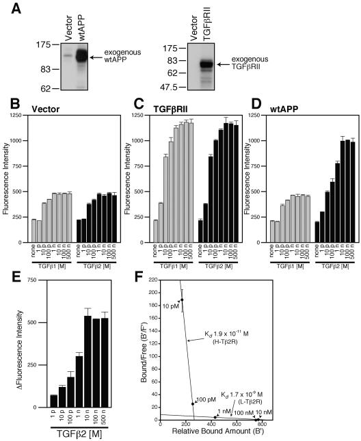
FIG. 1.
Overexpression of wtAPP induces the expression of TGFβ2-specific receptors. (A) F11 cells (7 × 10⁴ cells/well in 6-well plates) were transfected with 0.5 μg of the pcDNA3 vector, pcDNA3-TGFβRII, or pcDNA3-wtAPP. Cell lysates (10 μg in each lane) were immunoblotted with antibody to APP (22C11) or TGFβRII. (B to D) F11 cells (7 × 10⁴ cells/well in 6-well plates) were transfected with 0.25 μg of the pcDNA3 vector (B), pcDNA3-TGFβRII (C), or pcDNA3-wtAPP (D). At 36 h after transfection, the cells were combined with the indicated concentrations of TGFβ1 or TGFβ2. Immunofluorescence-based binding assays were performed as described in Materials and Methods. (E) The mean of immunofluorescence intensity numbers representing the association between TGFβ2 and F11 cells for each concentration of TGFβ2 shown in panel B were subtracted from immunofluorescence intensity numbers representing the association between TGFβ2 and F11 cells for the same concentration of TGFβ2 shown in panel D. Resulting intensity numbers were considered to correspond to the association between TGFβ2 and wtAPP-induced TGFβ2-specific receptors. (F) A simulated Scatchard plot of the association between TGFβ2 and wtAPP-induced TGFβ2-specific receptors based on the analysis shown in Table 1. A vertical bar for each point indicates an estimated range of bound/free numbers. p, pico; n, nano; B′, relative amount of bound TGFβ2; F′, estimated amount of free TGFβ2.

