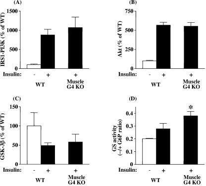
FIG. 5.
Insulin-stimulated IRS-1-associated PI3K (A), Akt (B), GSK3β (C), and glycogen synthase (D) activity levels in muscle from WT and muscle-G4KO mice. After an overnight fast, mice were injected i.v. with saline (−, white bars) or 10 U/kg insulin (+, black bars). Three min later, muscle was removed. (A) PI3K activities were measured in IRS-1 immunoprecipitates and were quantitated using a PhosphorImager. (B) Muscle lysates (500 μg) were subjected to immunoprecipitation with an Akt antibody that recognizes both Akt1 and Akt2. The immune pellets were assayed for kinase activity using Crosstide as the substrate. (C) Muscle lysates (500 μg) were subjected to immunoprecipitation with a GSK3β-specific antibody. The immune pellets were assayed for kinase activity using phospho-glycan synthase 1 as the substrate. (D) The ratio of glycogen synthase (GS) activity represents the activity measured in the absence divided by that in the presence of glucose-6-phosphate. Results are means ± SEM for four to six mice per group. *, P < 0.05 versus insulin-stimulated WT.

