Figure 5.
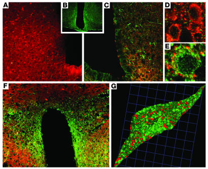
GM-CSF receptor immunohistochemistry. Synaptophysin (red) and GMRα immunofluorescence (green) were localized on neurons throughout mouse brain, including ARC and PVN. (A) Only synaptophysin immunofluorescence was observed in sections when antibody serum was preincubated with the immunizing peptide to block GMRα antibody binding. (B) A section stained with GMRα immunofluorescence alone. (C) GMRα immunofluorescence was colocalized with synaptophysin immunofluorescence in the ARC (low-magnification view). (D) High-magnification view of several synaptophysin-immunoreactive neurons that did not contain GMRα. (E) High-magnification view of GMRα-positive cells surrounded by synaptophysin immunofluorescence contacts in the ARC. (F) GMRα immunofluorescence was colocalized with synaptophysin immunofluorescence in the PVN (low-magnification view). (G) High-magnification 3-dimensional reconstruction of confocal images of a single neuron from the PVN, showing colocalization of GMRα and synaptophysin immunofluorescence. Sections are representative of 5 animals in which staining was examined.

