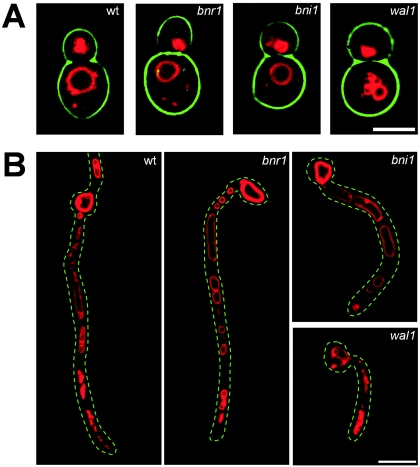
FIG. 2.
Analysis of endocytosis. Endocytosis and the vacuolar morphology of the indicated strains were analyzed by monitoring the uptake of FM4-64 during the yeast stage (A) and hyphal stage (B). In panel A, images of yeast cells that were grown overnight and subsequently stained with FM4-64 are shown. The wild type (wt) and formin mutants generate a single large vacuole. In contrast, the wal1 cells exhibit fragmented vacuoles. Movies 4 to 6 at the website http://pinguin.biologie.uni-jena.de/phytopathologie/pathogenepilze/index.html show the time course of uptake of FM4-64. In panel B, images of FM4-64-stained cells induced to form germ tubes (3 h at 37°C in the presence of serum) are shown. Wild-type and formin mutant germ tubes show the characteristic larger vacuoles in their terminal regions, while the wal1 mutant displays fragmented vacuoles throughout the initial germ tube. Scale bars, 5 μm (A) and 10 μm (B).

