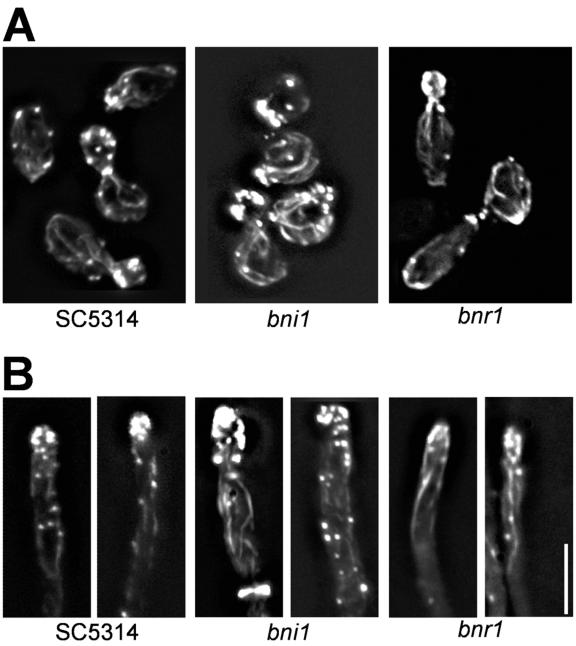
FIG. 5.
Analysis of the actin cytoskeleton. Images of rhodamine-phalloidin-stained cells of the indicated strains are shown. Cells were grown overnight in yeast extract-peptone-dextrose (YPD) at 30°C, inoculated into fresh YPD (A) or YPD plus 10% serum (B), and grown for 3 hours at 30°C (A) or 37°C (B) prior to fixation and staining. Bar, 10 μm.

