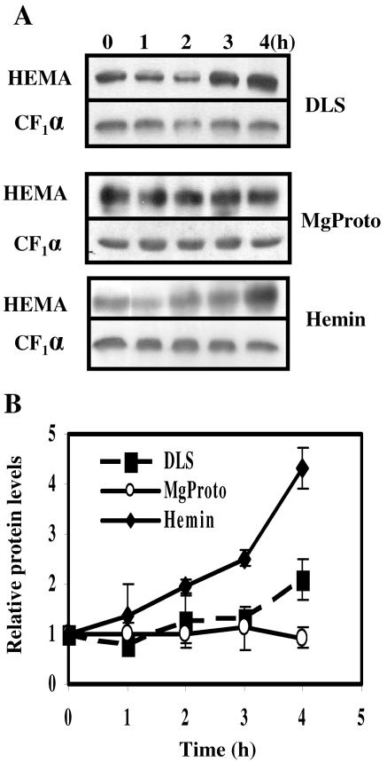
FIG. 5.
Changes in levels of GluTR after the application of light or tetrapyrroles (MgProto and hemin). (A) Soluble protein was extracted from wild-type cells preincubated in the dark for at least 20 h and treated with either hemin (final concentration, 4 μM) or MgProto (final concentration, 16 μM) in the dark or transferred from dark to light (fluence rate, 50 μmol m−2 s−1) (DLS). In each lane 50 μg of protein was separated by SDS-polyacrylamide gel electrophoresis and, after transfer to a membrane, immunodecorated with antibodies directed against C. reinhardtii GluTR. Signals were detected and quantified as described in Materials and Methods. Differences in the intensities of time zero signals are caused by different exposure times of the films. (B) Changes in GluTR levels in soluble protein from cells before and after a dark-to-light shift (DLS) or the addition of MgProto or hemin. The signals from at least three independent experiments were quantified and corrected for unequal loading by the respective signal for the α subunit of ATP synthase (CF1α). Values are given in arbitrary units relative to the continuous dark level (zero time point), which was set to 1. Error bars indicate standard errors.

