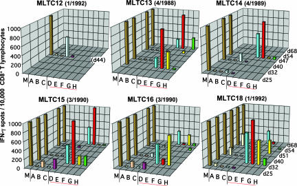
Fig. 4.
Specificity of tumor-reactive T cells enriched in DT MLTC. Independent autologous MLTC were performed with PBMC collected from patient DT during a 4-year period (Fig. 1). CD8+ MLTC responders were isolated and cryopreserved at various time points and later tested in 20-h IFN-γ ELISPOT assays for recognition of autologous melanoma cells (30,000 per well) (M) and COS-7 cells (20,000 per well) transiently transfected with expression plasmids encoding the antigen/HLA combinations tyrosinase/HLA-A*2601 (A), tyrosinase/HLA-B*3801 (B), gp100/HLA-B*07021 (C), SIRT2_v3mut/HLA-A*03011 (D), GPNMB_v2mut/HLA-A*03011 (E), SNRP116mut/HLA-A*03011 (F), RBAF600mut/HLA-B*07021 (G), SNRPD1mut/HLA-B*3801 (H). Data are means of duplicates and represent the number of spot forming cells per 15,000 (day 25) or 10,000 (later than day 25) CD8+ MLTC responders.

