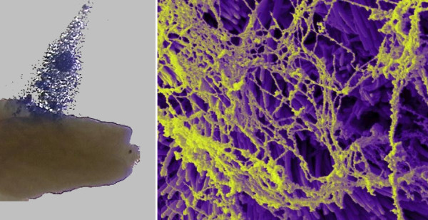
Figure 5.

Micrographs showing adhesion between the oocyte cumulus complex and the infundibulum. (A) Stereoscopic micrograph of an oocyte cumulus complex, colorized blue, being pulled off the surface of an infundibulum using forceps. The matrix of the complex adheres to the infundibulum. Complexes do not adhere to most other surfaces. (B) Scanning electron micrograph of cumulus matrix adhering to cilia on the outer surface of an infundibulum. The matrix was left behind by an oocyte cumulus complex that was picked-up by the infundibulum.
