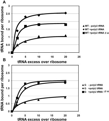
Figure 1.
Binding of deacylated tRNAPhe to ribosomes. The graph shows the number of tRNA molecules bound to wild-type ribosomes (A), or to ribosomes carrying the C2394G mutation (B), versus tRNA excess over ribosomes. The curves are marked according to the presence or absence of a polyU template and to the tRNA molecule used. Closed triangles indicate ribosomes with presence of tRNA and absence of polyU. Closed diamonds indicate ribosomes with presence of both tRNA and polyU. Closed squares correspond to ribosomes with presence of polyU and tRNA-Δ3′A. ‘tRNA’ corresponds to intact tRNAPhe, ‘tRNA-Δ3′A’ corresponds to tRNAPhe lacking the 3′-terminal adenine.

