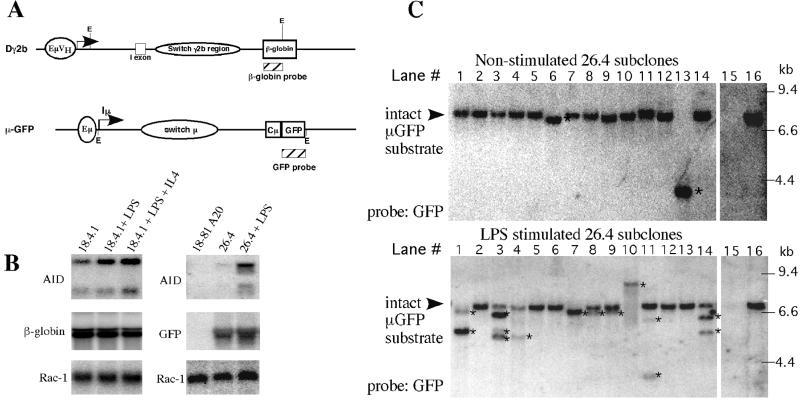
Figure 1.
Induction of intra-S region deletions detected in transfected ISR substrates. (A) Design of the Dγ2b and μ-GFP constructs showing the position of S region sequences in relation to restriction sites and hybridization probes (hatched boxes) used for Southern analysis. See text for details. E, EcoR1. (B) (Left) AID transcripts are detectable by Northern blotting in the Dγ2b-containing subclone 18.4.1 and increase after activation. Dγ2b transcripts as detected by the β-globin probe display constitutive expression from the substrate even after activation. Rac-1 levels were used to determine RNA loading (33). (Right) AID transcripts are induced in μ-GFP subclone 26.4 after activation with LPS. (Middle) A Northern blot with GFP probe showing constitutive expression of the μ-GFP construct in subclone 26.4. (Bottom) A Northern blot of the same RNA hybridized with a Rac-1 control probe to indicate RNA loading. (C) Deletions within the Sμ region of the μ-GFP construct occur after activation of 18–81 A20 cells with LPS. Southern blotting analysis on DNA from 18–81 A20 (lane 15), 26.4 (lane 16), and 26.4 subclones (lanes 1–14). μ-GFP transfected (26.4) 18–81 A20 cells were grown in LPS or media alone for 2 days and then subcloned at limiting dilution. Genomic DNA was isolated and digested with EcoR1 and screened with the GFP-specific probe shown in A. Rearrangements are denoted by *.

