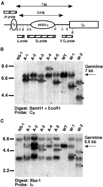
Figure 2.
Decreased frequency of Sμ deletions in hybridomas derived from AID−/− B cells compared with WT, IgM-secreting hybridomas. (A) Genomic organization of the JH-Cμ region of the IgH locus, including the location of the intronic IgH enhancer (Eμ), μS region (Sμ), and μ constant region exons (Cμ). B, BamH1; E, EcoR1; H, HindIII; S, Sac1; X, XbaI. Two-sided arrows indicate germ-line EcoR1/BamH1 and Xba1 restriction fragment sizes. Hatched boxes represent hybridization probes used for Southern blotting analyses. (B) Panel of hybridomas showing internal deletions within Sμ. Hybridoma DNA was doubly digested with BamH1/EcoR1 and Southern analysis was performed by using the 5′ Cμ−specific probe shown in A. W1 and W2 are two representative WT IgM-secreting hybridomas; A1-A5 are AID−/− hybridomas. NS-1 is the hybridoma fusion partner. Kidney DNA (WT) indicates germ-line configuration. (C) To confirm that the deletions were between the Eμ and Cμ regions, Southern blotting analysis was done on Xba1-digested DNA and probed with the Iμ probe shown in A. Results on all Sμ-deleting hybridomas are summarized in Table 2. Rearrangements are denoted by *.

