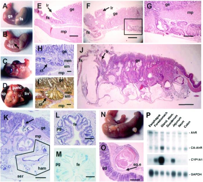
Figure 3.
Neoplastic lesions and intestinal metaplasia in the stomach of CA-AhR mice. (A) Normal stomach from a 12-month-old wild-type male showing the forestomach (fs) and glandular stomach (gs). (B) At 3–4 months of age single small cysts close to the limiting ridge were seen in CA-AhR mice (arrow). (C) In older CA-AhR animals (6–12 months old) the number of cystic tumors increased and occupied a larger area of the stomach. (D) In the most severe cases (9–12 months of age), the stomach was adherent to adjacent organs such as spleen (sp), pancreas (panc), and liver (liv). (E) Normal stomach from a 6-month-old wild-type male mouse showing the muscularis propria layer (mp) and limiting ridge (lr) constituting the border between the squamous epithelium of the forestomach and the glandular epithelium (ge, HE staining). (Bar, 0.5 mm.) (F) Close to the limiting ridge a rupture of the submucosa by neoplastic crypts is seen in a 3.5-month-old CA-AhR male. Note glands within the stroma of the limiting ridge (HE). (Bar, 0.5 mm.) (G) Larger magnification of the boxed area in F (HE). (Bar, 0.15 mm.) (H and I) Invading crypts surrounded by connective tissue (ct) invade the submucosa (sm) by penetrating through the muscularis mucosa (mm) and submucosa layers and further into the muscularis propria in a 9-month-old CA-AhR female (H, HE staining; I, van Gieson staining). (Bars, 0.1 mm.) (J) Stomach from a 12-month-old CA-AhR male with severely distorted tissue architecture (HE). (Bar, 1.25 mm.) (K) Glands underlying the serosa (ser) in a 12-month-old CA-AhR female with characteristics of a hamartoma (ham), i.e., lymphatic tissue, vessels, and fat (HE). (Bar, 0.5 mm.) Note also invasion (arrow) of glands from the glandular epithelium into the muscularis propria. (L and M) Glandular structures located in the muscularis propria with cells resembling foveolar epithelium (fe) and pyloric glands (pg) showing intestinal metaplasia in a 9-month-old CA-AhR male (L, HE staining; M, Alcian blue pH 2.5 staining). (Bars, 0.1 mm.) (N and O) Squamous cysts on the caecum showing colonic glands (cg) and squamous epithelium (sq.e) of a 9-month-old CA-AhR male (HE). (Bar, 0.5 mm.) (P) The expression and activity of CA-AhR in the gastrointestinal tract is highest in the glandular stomach. RNA blot showing expression of CA-AhR, endogenous AhR (AhR), CYP1A1, and glyceraldehyde-3-phosphate dehydrogenase (GAPDH) mRNA in different parts of the alimentary tract of homozygous CA-AhR mice 3 months of age.

