Figure 3.
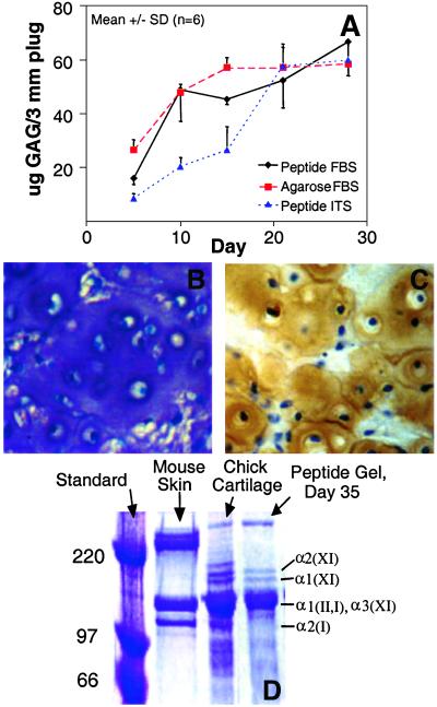
Matrix accumulation in chondrocyte-seeded peptide hydrogel. (A) Total GAG accumulation in cell-seeded peptide hydrogel cultured in FBS and ITS/FBS medium and in cell-seeded agarose. (B) Toluidine blue staining of chondrocyte-seeded peptide hydrogel cultured in 10% FBS, day 15. (C) Immunohistochemical staining for type II collagen in cell-seeded peptide hydrogel cultured in 10% FBS, day 15. Image width for B and C = 175 μm. (D) SDS/PAGE of collagens extracted from day 35 samples of chondrocyte-seeded peptide hydrogel cultured in 1% ITS with 0.2% FBS. Standards: Chick cartilage for collagen II and XI banding pattern. Mouse skin identifies collagen I α-helix 2, indicative of collagen expression of a dedifferentiated, fibroblastic phenotype.

