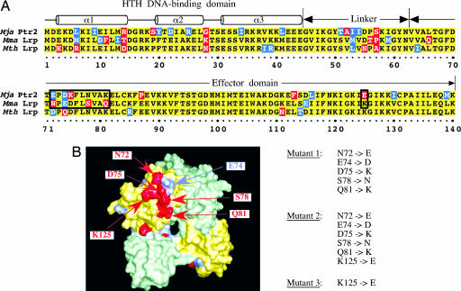
Fig. 1.
Two methanococcal homologues of Ptr2. (A) Sequence alignment of Mja Ptr2, Mth Lrp, and Mma Lrp. Conserved, similar, and differing residues are shaded in yellow, blue, and red, respectively. The putative DNA-binding, linker, and effector domains are indicated above the alignment. (B) Homology model of Mma Lrp, shown in surface representation as a dimer of green and yellow protomers. Positions of sequence divergence from Mja Ptr2 are colored as in A, with putative activation determinants labeled. Also shown are the amino acid substitutions that generate Mma Lrp mutants 1, 2, and 3.

