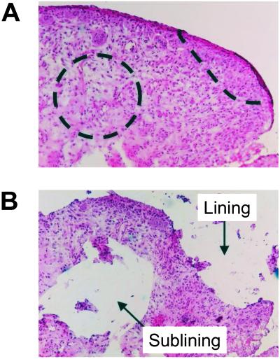
Figure 1.
Microdissection of RA synovial tissues. Frozen synovial tissues were cut into 10-μm sections. Intimal lining and sublining regions were microdissected by using a sterile blade under light microscopy. An example of the size and location of microdissected regions is shown (A: before microdissection, B: after microdissection). The area of each region was ≈0.25 mm2, and the number of the cells in the region was estimated at approximately 1,000 cells. (Magnification: ×100.)

