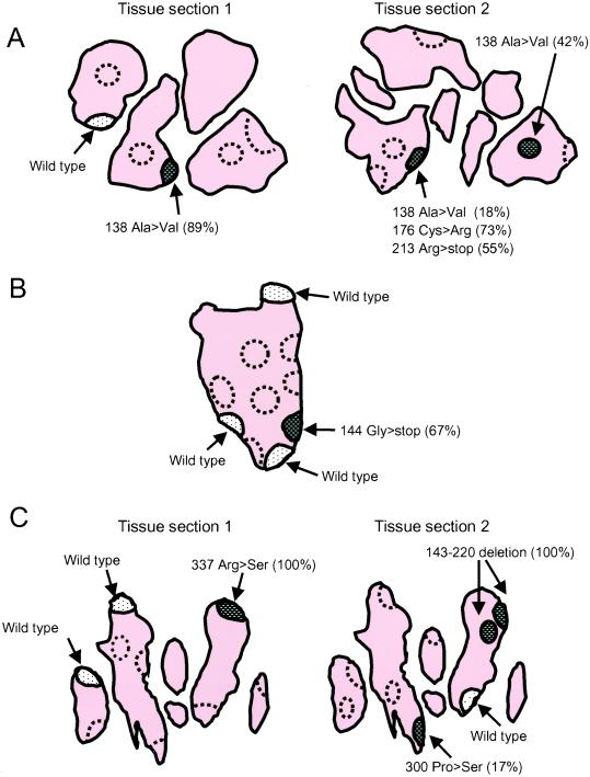
Figure 4.
Localization of p53 mutations in RA synovial tissues. Several schematic drawings of frozen tissue sections are shown to illustrate the location of p53 expression and mutations. The outer edge of the section indicates lining regions, and interior is sublining. Note that folds in the tissue result in the appearance of several discrete pieces of tissue from a single block. The regions shown by dashed lines contained no detectable p53 mRNA by a nested PCR. The predominant p53 sequences are show for each p53 mRNA-positive region. (A) Patient RA1. Six regions (three lining regions and three sublining regions) were microdissected in the first section (tissue section 1). p53 mRNA was detected in two of three lining regions, whereas none of the three sublining regions contained detectable p53 mRNA. In one lining region (hatched area), 89% of the p53 subclones contained codon 138 Ala (GCC) to Val (GTC) missense mutation. Many of the mutant subclones contained more than one mutation, suggesting sequential accumulation of mutations by various cells. In contrast, most of the p53 subclones contained wild-type p53 in the other region (dotted area). In an adjacent section from the same tissue block (tissue section 2), the codon 138 Ala to Val mutation was also detected. Additional mutations were also abundant in the same region (codon 176 Cys>Arg, codon 213 Arg>stop). (B) Patient RA2. Ten regions (seven lining regions and three sublining regions) were microdissected. p53 mRNA was detected in four of seven lining regions; however, none of the three sublining regions contained detectable p53 mRNA. In one lining region (hatched area), 67% of p53 subclones contained the codon 144 Gly (CAG) to stop codon (TAG) mutation, whereas the remaining three lining regions (dotted area) were primarily wild-type p53. (C) Patient RA8. Nine regions (eight lining and one sublining) were microdissected. p53 mRNA was detected in three of eight lining regions, but not in the sublining. In one lining region (hatched area), 100% of p53 subclones contained the codon 337 Arg (CGC) to Ser (AGC) mutation, whereas the remaining two lining regions (dotted area) were primarily wild-type p53.

