
Figure 1.
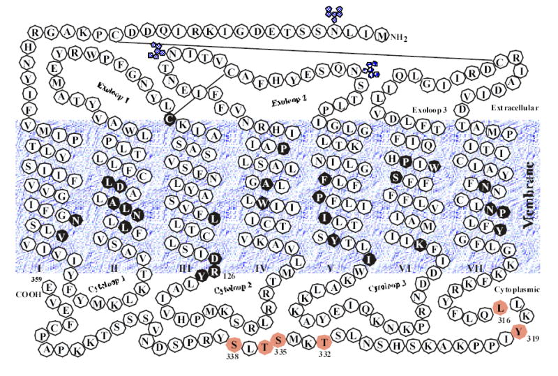
The secondary structure model of the rat AT1 angiotensin II receptor. Putative -helical transmembrane domains I–VII, three potential glycosylation sites, and two disulfide bonds are shown. Residues and motifs in closed black circles are highly conserved in more than 90% of GPCRs. The amino acids in the cytoplasmic tail of the receptor that have been suggested to be important for receptor internalization are shaded. The membrane interface boundaries for all seven TM helices are tentative.