Figure 5.
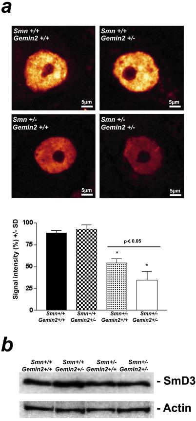
Sm protein immunoreactivity in the nucleus of spinal motoneurons of 5-month-old Gemin2+/−, Smn+/−, Gemin2+/−/Smn+/−, and wild-type mice. (a) The monoclonal anti-Sm protein antibody Y12 was used for immunohistochemical analysis of the Sm protein distribution in the nucleus of spinal motoneurons of 5-month-old Gemin2+/−, Smn+/−, Gemin2+/−/Smn+/−, and wild-type mice. A significant reduction of Sm proteins in the nucleus of spinal motoneurons was detectable in Smn/Gemin2 double heterozygous deficient mice (*) in comparison to wild type, Gemin2+/−, and Smn+/− motoneurons. Distribution of the Sm proteins within the cell body of each genotype was not altered. (Lower) The semiquantitative analysis of Sm protein levels in spinal motoneuron nuclei. Reduced Sm protein signal intensity was measured in spinal motoneurons of Smn+/− (−39%, *) and even more (P < 0.05) in Smn+/−/Gemin2+/− mice in comparison to wild-type and Gemin2+/− mice. (b) Similar signal intensities for SmD3 proteins in spinal cord of wild-type, Gemin2+/−, Smn+/−, and Smn/Gemin2 double heterozygous-deficient mice were observed. The anti-actin antibody was used as a control showing equal protein concentration in each lane.

