
Figure 6.
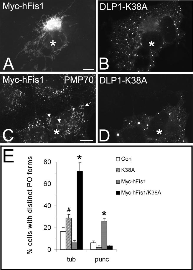
Inhibition of DLP1 function interferes with peroxisomal fission induced by hFis1 expression. COS-7 cells were cotransfected with Myc-hFis1 and GFP-DLP1-K38A (A–D) and immunostained with antibodies to the Myc tag of hFis1 (A) or to PMP70 (C). The corresponding GFP fluorescence of DLP1-K38A is shown in B and D. Note the pronounced elongation of peroxisomes in DLP1-K38A coexpressing cells (arrows). A quantitative analysis of peroxisome morphology is shown in E. Cells were categorized as cells with tubular (tub) or punctiform (punc) peroxisomes (percentage of total) (see Materials and Methods); *p < 0.01, # p < 0.05 compared with controls. Con, untransfected control. Asterisks, coexpressing cells. N, nucleus. Bars, 10 μm.