Figure 5.
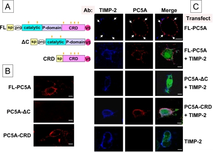
Cell surface colocalization of TIMP-2 and FL-PC5A. (A) Schematic representation of the 3 constructs used for the analysis by confocal microscopy. (B) Cell surface labeling of COS-1 cells expressing FL-PC5A, PC5A-ΔC, or PC5A-CRD with Ab:V5. (C) Cell surface immunofluorescence of COS-1 cells probed with Ab:V5 and Ab:TIMP-2. TIMP-2 is in a phCMV vector and PC5A and its derivatives (all tagged with a V5 epitope) in the pIRES2-EGFP vector. Cells were transfected with either FL-PC5A and the empty phCMV vector, or with TIMP-2 plus either FL-PC5A, PC5A-ΔC or PC5A-CRD, or the pIRES2-EGFP vector alone. The green-EGFP fluorescence is a control of PC5-transfection, whereas the blue and red labeling are indicating TIMP-2 and the various forms of PC5A, respectively. Colocalization areas are seen in pink. Arrows point to colocalization areas of endogenous TIMP-2 and transfected FL-PC5A. Bars, 10 μm.

