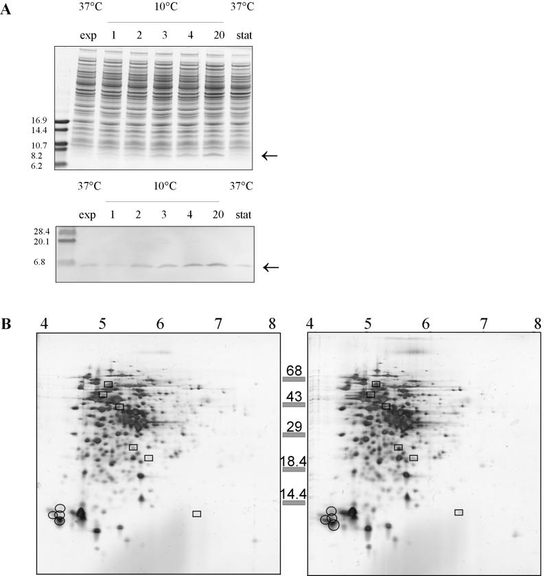
FIG. 2.
Analysis of L. monocytogenes LO28 proteins. (A) Upper panel, one-dimensional gel electrophoresis of mid-exponential-phase cells (exp) (37°C), cells cold shocked at 10°C for 1, 2, 3, 4, and 20 h, and cells in the stationary phase (stat) at 37°C; lower panel, Western blot of an identical one-dimensional gel with anti-CspB from B. subtilis. (B) 2D-E of cell extracts of L. monocytogenes LO28 with pI values ranging from 3 to 10. Proteins induced at least threefold are enclosed in boxes, and the CSPs are circled. Left panel, exponential-phase cells at 37°C; right panel, cells 4 h after cold shock at 10°C.

