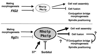
FIG. 7.
Model of FIG2 function in mating morphogenesis, cell fusion, and cell integrity. In wild-type cells, Fig2p is deposited in the cell wall during and throughout mating cell morphogenesis. Polarized growth activates Rho1/Pkc1 signaling, increasing cell wall assembly in the projection area and delaying cell fusion until a partner is sensed. Once activated Rho1/Pkc1 levels fall and cell fusion begins, lateral expansion of the conjugation bridge and subsequent spindle positioning occurs. In fig2Δ cells, Fig2p is absent from the mating cell wall, leading to its disorganization or perhaps increased pressure in the region of the projection where Fig2p is normally present. This change is signaled, leading to more elevated Rho1/Pkc1 activity. This higher level of activity normally feeds back to enhance cell wall biogenesis, partially compensating for absence of Fig2p. Elevated activity nonetheless leads to delays in cell fusion and spindle position and may delay lateral morphogenesis of the conjugation bridge. Conditions of higher temperature or sorbitol support enhance or suppress these phenotypes, respectively, through their effects on Rho1/Pkc1 signaling.

