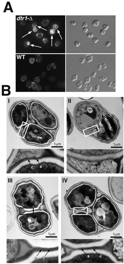
FIG. 4.
Morphological spore wall defects associated with the deletion of DTR1. (A) Fluorescence microscopy with Calcofluor white of dtr1-Δ and wild-type asci. The deletion of DTR1 did not result in a uniform phenotype, instead we observed a mixture of unstained, morphologically wild-type asci and asci containing spores whose chitosan layer became accessible for the fluorescence dye (marked with arrows). Electron microscopy further confirmed the heterogeneity of dtr1-Δ asci. (B) Electron micrographs of representative examples of dtr1-Δ asci. (I) An ascus with the typical appearance of the wild-type spore wall. The dityrosine-containing surface layer (s) is very electron dense and is clearly distinguishable from the underlying chitosan layer (c). 25% of the dtr1-Δ asci showed an unaltered spore wall structure. (II) An ascus containing one spore with a wild-type appearance and one spore lacking the surface layer. Approximately 15% of all asci contained one or more spores with this phenotype. (III and IV) Two examples of asci with an altered surface structure. The surface layer was present but less electron dense than in the wild type and hardly discernible from the chitosan layer. Approximately 60% of the dtr1-Δ asci showed this phenotype. The rectangles indicate the areas shown in the insets at a fourfold magnification.

