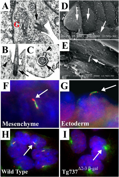
Figure 1. Cilia Are Present on Both Mesenchymal and Ectodermal Cells of the Developing Limb.
(A–C) Transmission electron micrographs of limb bud mesenchyme show cilia (arrows) closely associated with the Golgi apparatus (“G”). The cilia exhibited a 9 + 0 structure (C) and are often found in deep depressions in the membranes (B). Frequently, small vesicles are observed fusing or budding with the surrounding membrane (arrowheads in [B] and [C]).
(D and E) Scanning electron micrographs of the limb ectoderm show a single cilium (arrows) on nearly all ectodermal cells.
(F and G) Immunolocalization of Polaris (red) and acetylated α-tubulin (green) in frozen sections of limb buds shows that Polaris concentrates at the base and tip of the axoneme in both mesenchymal (F) and ectodermal (G) cells. Nuclei are blue.
(H and I) In primary cultures of cells isolated from E11.5 limb buds, cilia (arrow in H) are also present when visualized with anti-acetylated α-tubulin (green) and anti-Polaris (red) antisera (H). Cilia are absent on cells isolated from Tg737 Δ2–3β-gal mutant limb buds (I); however, the stabilized microtubules were still evident around the basal body region (arrow). The nuclear staining for Polaris is present in the Tg737 Δ2–3β-gal cells, indicating that it is nonspecific. Nuclei are blue.

