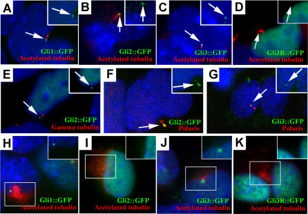
Figure 5. GFP-Tagged Gli Proteins Localize to the Distal Tip of the Cilium in Primary Limb Cell Cultures.
(A–D) Cells were isolated from limb buds of wild-type embryos at E11.5 and infected with the indicated adenovirus. All three full-length GFP-tagged Gli proteins (green) localize to a domain in the cilium axoneme, which is visualized with anti-acetylated α-tubulin staining (red). In contrast, Gli3R::GFP is restricted to the nucleus and is not detected in this domain (D).
(E) The full-length Gli::GFP proteins (Gli2::GFP shown here) do not colocalize with the basal body at the base of the cilium, which is visualized with anti-γ-tubulin staining (red), indicating that the full-length Gli proteins localize to the tips of the cilia.
(F and G) Gli2::GFP (F) and Gli3::GFP (G) colocalize with a subdomain of Polaris (red) at the distal tip of the cilium.
(H–K) In Tg737 Δ2–3β-gal mutant limb bud cells, the GFP-tagged Gli1 (H), Gli2 (I), and Gli3 (J) proteins localize to the nucleus and in the region of stabilized microtubules around the MTOC marked by anti-acetylated α-tubulin. In contrast, the processed form of Gli3 (Gli3R::GFP) (K) is detected only in the nucleus.
Insets in all panels show the GFP (green) and nuclear (blue) staining only for the indicated cilium (arrow) or region (box).

