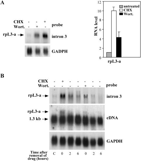
Figure 2.
Expression of alternatively spliced mRNA of the rpL3 gene (rpL3-a mRNA). (A) Left panel: northern blots of total RNA from untreated Calu-6 cells and from the same cells after incubation with either CHX or Wort. The probe used for hybridization is specific for the portion of intron retained by alternative splicing. Right panel: rpL3-a mRNA was quantified by PhosphorImager, normalized to GAPDH levels and expressed in a graph as a ratio to endogenous levels in untreated cells. Numerical values are the average of three independent experiments, reported with SDs. (B) Decay of the drug-stabilized rpL3-a mRNA. Total RNA was isolated from Calu-6 cells before treatment (lane C), after treatment with CHX or with Wort., and 2 and 6 h after drug removal. Northern blot was performed with the indicated probes.

